Question
Question asked by Filo student

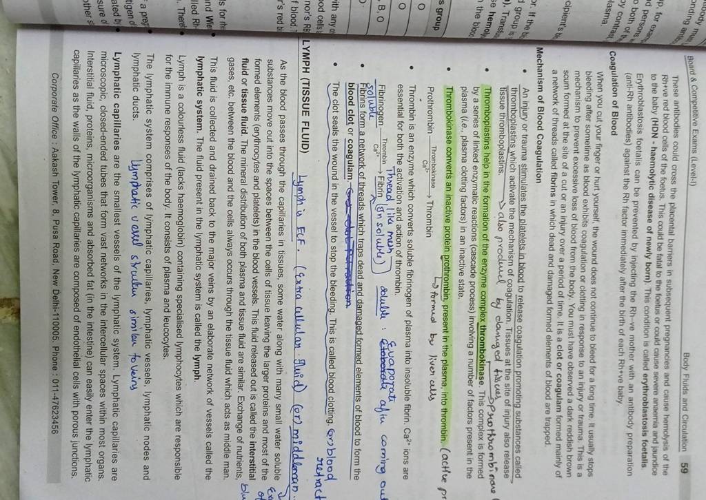
Board Competitive Exams (Level-1) Body Fluids and Circulation 59 These antibodies could cross the placental barriers in subsequent pregnancies and cause hemolysis of the Rh+ve red blood cells of the foetus. This could be fatal to the foetus or could cause severe anaemia and jaundice to the baby (HDN - haemolytic disease of newly born). This condition is called erythroblastosis foetalis. Erythroblastosis foetalis can be prevented by injecting the Rh-ve mother with an antibody preparation (anti-Rh antibodies) against the Rh factor immediately after the birth of each Rh+ve baby. Coagulation of Blood When you cut your finger or hurt yourself, the wound does not continue to bleed for a long time, It usually stops bleeding after sometime as blood exhibits coagulation or clotting in response to an injury or trauma. This is a mechanism to prevent excessive loss of blood from the body. You must have observed a dark reddish brown scum formed at the site of a cut or an injury over a period of time. It is a clot or coagulam formed mainly of a network of threads called fibrins in which dead and damaged formed elements of blood are trapped. Mechanism of Blood Coagulation - An injury or trauma stimulates the platelets in blood to release coagulation promoting substances called thromboplastins which activate the mechanism of coagulation. Tissues at the site of injury also release tissue thromboplastins. also producel by daniged tissces - Thromboplastins help in the formation of the enzyme complex thrombokinase. This prothro mbindre by a series of linked enzymatic reactions (cascade process) involving a plasma (i.e., plasma clotting factors) in an inactive state. - Thrombokinase converts an inactive protein prothrombin, present in the plasma, into thrombin. (active pr Prothrombin Thrombin - Thrombin is an enzyme which converts soluble fibrinogen of plasma into insoluble fibrin. ions are essential for both the activation and action of thrombin. - Fibrins form a network of threads which traps dead and damaged formed elements of blood to form the blood clot or coagulam. - The clot seals the wound in the vessel to stop the bleeding. This is called blood clotting. Groblood retract LYMPH (TISSUE FLUID) Lymph is ECF. (Extra ullular. Fluid) (or) middlonan. As the blood passes through the capillaries in tissues, some water along with many small water soluble substances move out into the spaces between the cells of tissue leaving the larger proteins and most of the formed elements (erythrocytes and platelets) in the blood vessels. This fluid released out is called the interstitial fluid or tissue fluid. The mineral distribution of both plasma and tissue fluid are similar. Exchange of nutrients, gases, etc. between the blood and the cells always occurs through the tissue fluid which acts as middle man. This fluid is collected and drained back to the major veins by an elaborate network of vessels called the lymphatic system. The fluid present in the lymphatic system is called the lymph. Lymph is a colourless fluid (lacks haemoglobin) containing specialised lymphocytes which are responsible for the immune responses of the body. It consists of plasma and leucocytes. The lymphatic system comprises of lymphatic capillaries, lymphatic vessels, lymphatic nodes and lymphatic ducts. Lymphatic uesel struetur similan foveins Lymphatic capillaries are the smallest vessels of the lymphatic system. Lymphatic capillaries are microscopic, closed-ended tubes that form vast networks in the intercellular spaces within most organs. Interstitial fluid, proteins, microorganisms and absorbed fat (in the intestine) can easily enter the lymphatic capillaries as the walls of the lymphatic capillaries are composed of endothelial cells with porous junctions. Corporate Office : Aakash Tower, 8. Pusa Road, New Delhi-110005. Phone : 011-47623456
Found 3 tutors discussing this question
Discuss this question LIVE
15 mins ago

One destination to cover all your homework and assignment needs
Learn Practice Revision Succeed

Instant 1:1 help, 24x7
60, 000+ Expert tutors

Textbook solutions
Big idea maths, McGraw-Hill Education etc

Essay review
Get expert feedback on your essay

Schedule classes
High dosage tutoring from Dedicated 3 experts
Practice more questions on Human Physiology
Question 1
Easy
Views: 5,871
Question 4
Easy
Views: 6,017
Students who ask this question also asked
Question 1
Views: 5,832
Question 2
Views: 5,730
Question 4
Views: 5,892


Stuck on the question or explanation?
Connect with our Biology tutors online and get step by step solution of this question.
231 students are taking LIVE classes
| Question Text | Board Competitive Exams (Level-1)
Body Fluids and Circulation 59
These antibodies could cross the placental barriers in subsequent pregnancies and cause hemolysis of the Rh+ve red blood cells of the foetus. This could be fatal to the foetus or could cause severe anaemia and jaundice to the baby (HDN - haemolytic disease of newly born). This condition is called erythroblastosis foetalis.
Erythroblastosis foetalis can be prevented by injecting the Rh-ve mother with an antibody preparation (anti-Rh antibodies) against the Rh factor immediately after the birth of each Rh+ve baby.
Coagulation of Blood
When you cut your finger or hurt yourself, the wound does not continue to bleed for a long time, It usually stops bleeding after sometime as blood exhibits coagulation or clotting in response to an injury or trauma. This is a mechanism to prevent excessive loss of blood from the body. You must have observed a dark reddish brown scum formed at the site of a cut or an injury over a period of time. It is a clot or coagulam formed mainly of a network of threads called fibrins in which dead and damaged formed elements of blood are trapped.
Mechanism of Blood Coagulation
- An injury or trauma stimulates the platelets in blood to release coagulation promoting substances called thromboplastins which activate the mechanism of coagulation. Tissues at the site of injury also release tissue thromboplastins. also producel by daniged tissces
- Thromboplastins help in the formation of the enzyme complex thrombokinase. This prothro mbindre by a series of linked enzymatic reactions (cascade process) involving a plasma (i.e., plasma clotting factors) in an inactive state.
- Thrombokinase converts an inactive protein prothrombin, present in the plasma, into thrombin. (active pr Prothrombin Thrombin
- Thrombin is an enzyme which converts soluble fibrinogen of plasma into insoluble fibrin. ions are essential for both the activation and action of thrombin.
- Fibrins form a network of threads which traps dead and damaged formed elements of blood to form the blood clot or coagulam.
- The clot seals the wound in the vessel to stop the bleeding. This is called blood clotting. Groblood retract
LYMPH (TISSUE FLUID) Lymph is ECF. (Extra ullular. Fluid) (or) middlonan.
As the blood passes through the capillaries in tissues, some water along with many small water soluble substances move out into the spaces between the cells of tissue leaving the larger proteins and most of the formed elements (erythrocytes and platelets) in the blood vessels. This fluid released out is called the interstitial fluid or tissue fluid. The mineral distribution of both plasma and tissue fluid are similar. Exchange of nutrients, gases, etc. between the blood and the cells always occurs through the tissue fluid which acts as middle man.
This fluid is collected and drained back to the major veins by an elaborate network of vessels called the lymphatic system. The fluid present in the lymphatic system is called the lymph.
Lymph is a colourless fluid (lacks haemoglobin) containing specialised lymphocytes which are responsible for the immune responses of the body. It consists of plasma and leucocytes.
The lymphatic system comprises of lymphatic capillaries, lymphatic vessels, lymphatic nodes and lymphatic ducts.
Lymphatic uesel struetur similan foveins
Lymphatic capillaries are the smallest vessels of the lymphatic system. Lymphatic capillaries are microscopic, closed-ended tubes that form vast networks in the intercellular spaces within most organs. Interstitial fluid, proteins, microorganisms and absorbed fat (in the intestine) can easily enter the lymphatic capillaries as the walls of the lymphatic capillaries are composed of endothelial cells with porous junctions.
Corporate Office : Aakash Tower, 8. Pusa Road, New Delhi-110005. Phone : 011-47623456 |
| Updated On | Nov 30, 2022 |
| Topic | Human Physiology |
| Subject | Biology |
| Class | Class 11 |
| Answer Type | Video solution: 1 |
| Upvotes | 58 |
| Avg. Video Duration | 12 min |


